In cell biology, mitosis is a part of the cell cycle when replicated chromosomes are separated into two new nuclei. In general, mitosis (division of the nucleus) is preceded by the S stage of interphase (during which the DNA is replicated) and is often accompanied or followed by cytokinesis, which divides the cytoplasm, organelles and cell membrane into two new cells containing roughly equal shares of these cellular components.[1] Mitosis and cytokinesis together define the mitotic (M) phase of an animal cell cycle—the division of the mother cell into two daughter cells genetically identical to each other.
The process of mitosis is divided into stages corresponding to the completion of one set of activities and the start of the next. These stages are prophase, prometaphase, metaphase, anaphase, and telophase. During mitosis, the chromosomes, which have already duplicated, condense and attach to spindle fibers that pull one copy of each chromosome to opposite sides of the cell.[2] The result is two genetically identical daughter nuclei. The rest of the cell may then continue to divide by cytokinesis to produce two daughter cells.[3] Producing three or more daughter cells instead of normal two is a mitotic error called tripolar mitosis or multipolar mitosis (direct cell triplication / multiplication).[4] Other errors during mitosis can induce apoptosis (programmed cell death) or cause mutations. Certain types of cancer can arise from such mutations.[5]
Mitosis occurs only in eukaryotic cells. Prokaryotic cells, which lack a nucleus, divide by a different process called binary fission. Mitosis varies between organisms.[6] For example, animal cells undergo an "open" mitosis, where the nuclear envelope breaks down before the chromosomes separate, whereas fungi undergo a "closed" mitosis, where chromosomes divide within an intact cell nucleus.[7] Most animal cells undergo a shape change, known as mitotic cell rounding, to adopt a near spherical morphology at the start of mitosis. Most human cells are produced by mitotic cell division. Important exceptions include the gametes – sperm and egg cells – which are produced by meiosis.
Protein synthesis is the process whereby biological cells generate new proteins; it is balanced by the loss of cellular proteins via degradation or export. Translation, the assembly of amino acids by ribosomes, is an essential part of the biosynthetic pathway, along with generation of messenger RNA (mRNA), aminoacylation of transfer RNA (tRNA), co-translational transport, and post-translational modification. Protein biosynthesis is strictly regulated at multiple steps.[1]They are principally during transcription (phenomena of RNA synthesis from DNA template) and translation (phenomena of amino acid assembly from RNA).
The cistron DNA is transcribed into the first of a series of RNA intermediates. The last version is used as a template in synthesis of a polypeptide chain. Protein will often be synthesized directly from genes by translatingmRNA. However, when a protein must be available on short notice or in large quantities, a protein precursor is produced. A proprotein is an inactive protein containing one or more inhibitory peptides that can be activated when the inhibitory sequence is removed by proteolysis during posttranslational modification. A preprotein is a form that contains a signal sequence (an N-terminal signal peptide) that specifies its insertion into or through membranes, i.e., targets them for secretion.[2] The signal peptide is cleaved off in the endoplasmic reticulum.[2]Preproproteins have both sequences (inhibitory and signal) still present.
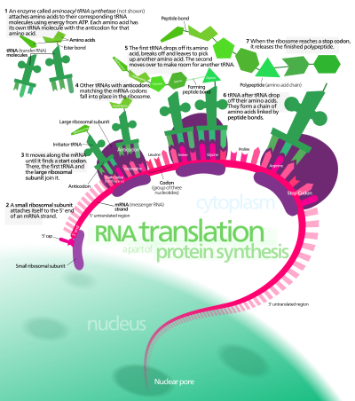
In protein synthesis, a succession of tRNA molecules charged with appropriate amino acids are brought together with an mRNA molecule and matched up by base-pairing through the anti-codons of the tRNA with successive codons of the mRNA. The amino acids are then linked together to extend the growing protein chain, and the tRNAs, no longer carrying amino acids, are released. This whole complex of processes is carried out by the ribosome, formed of two main chains of RNA, called ribosomal RNA (rRNA), and more than 50 different proteins. The ribosome latches onto the end of an mRNA molecule and moves along it, capturing loaded tRNA molecules and joining together their amino acids to form a new protein chain.[3]
Protein biosynthesis, although very similar, is different for prokaryotes and eukaryotes.
TRANSCRIPTION
In transcription an mRNA chain is generated, with one strand of the DNA double helix in the genome as a template. This strand is called the template strand. Transcription can be divided into 3 stages: initiation, elongation, and termination, each regulated by a large number of proteins such as transcription factors and coactivators that ensure that the correct gene is transcribed.
Transcription occurs in the cell nucleus, where the DNA is held. The DNA structure of the cell is made up of two helixes made up of sugar and phosphate held together by hydrogen bonds between the bases of opposite strands. The sugar and the phosphate in each strand are joined together by stronger phosphodiester covalent bonds. The DNA is "unzipped" (disruption of hydrogen bonds between different single strands) by the enzyme helicase, leaving the single nucleotide chain open to be copied. RNA polymerase reads the DNA strand from the 3-prime (3') end to the 5-prime (5') end, while it synthesizes a single strand of messenger RNA in the 5'-to-3' direction. The general RNA structure is very similar to the DNA structure, but in RNA the nucleotide uracil takes the place that thymine occupies in DNA. The single strand of mRNA leaves the nucleus through nuclear pores, and migrates into the cytoplasm.
The first product of transcription differs in prokaryotic cells from that of eukaryotic cells, as in prokaryotic cells the product is mRNA, which needs no post-transcriptional modification, whereas, in eukaryotic cells, the first product is called primary transcript, that needs post-transcriptional modification (capping with 7-methyl-guanosine, tailing with a poly A tail) to give hnRNA (heterogeneous nuclear RNA). hnRNA then undergoes splicing of introns (noncoding parts of the gene) via spliceosomes to produce the final mRNA.
TRANSLATION
The synthesis of proteins from RNA is known as translation. In eukaryotes, translation occurs in the cytoplasm, where the ribosomes are located. Ribosomes are made of a small and large subunit that surround the mRNA. In translation, messenger RNA (mRNA) is decoded to produce a specific polypeptide according to the rules specified by the trinucleotide genetic code. This uses an mRNA sequence as a template to guide the synthesis of a chain of amino acids that form a protein. Translation proceeds in four phases: activation, initiation, elongation, and termination (all describing the growth of the amino acid chain, or polypeptide that is the product of translation).
In activation, the correct amino acid (AA) is joined to the correct transfer RNA (tRNA). While this is not, in the technical sense, a step in translation, it is required for translation to proceed. The AA is joined by its carboxyl group to the 3' OH of the tRNA by an ester bond. When the tRNA has an amino acid linked to it, it is termed "charged". Initiation involves the small subunit of the ribosome binding to 5' end of mRNA with the help of initiation factors (IF), other proteins that assist the process. Elongation occurs when the next aminoacyl-tRNA (charged tRNA) in line binds to the ribosome along with GTP and an elongation factor. Termination of the polypeptide happens when the A site of the ribosome faces a stop codon (UAA, UAG, or UGA). When this happens, no tRNA can recognize it, but releasing factor can recognize nonsense codons and causes the release of the polypeptide chain. The capacity of disabling or inhibiting translation in protein biosynthesis is used by some antibiotics such as anisomycin, cycloheximide, chloramphenicol, tetracycline, streptomycin, erythromycin, puromycin,
DNA Structure and Replication: Crash Course Biology #10
In molecular biology, DNA replication is the biological process of producing two identical replicas of DNA from one original DNA molecule. This process occurs in all living organisms and is the basis for biological inheritance. DNA is made up of a double helix of two complementary strands. During replication, these strands are separated. Each strand of the original DNA molecule then serves as a template for the production of its counterpart, a process referred to as semiconservative replication. Cellular proofreading and error-checking mechanisms ensure near perfect fidelity for DNA replication.[1][2]
In a cell, DNA replication begins at specific locations, or origins of replication, in the genome.[3] Unwinding of DNA at the origin and synthesis of new strands results in replication forks growing bi-directionally from the origin. A number of proteins are associated with the replication fork to help in the initiation and continuation of DNA synthesis. Most prominently, DNA polymerase synthesizes the new strands by adding nucleotides that complement each (template) strand. DNA replication occurs during the S-stage of interphase.
DNA replication can also be performed in vitro (artificially, outside a cell). DNA polymerases isolated from cells and artificial DNA primers can be used to initiate DNA synthesis at known sequences in a template DNA molecule. The polymerase chain reaction (PCR), a common laboratory technique, cyclically applies such artificial synthesis to amplify a specific target DNA fragment from a pool of DNA.
By I, Madprime, CC BY-SA 3.0, https://commons.wikimedia.org/w/index.php?curid=2497221
DNA usually exists as a double-stranded structure, with both strands coiled together to form the characteristic double-helix. Each single strand of DNA is a chain of four types of nucleotides. Nucleotides in DNA contain a deoxyribose sugar, a phosphate, and a nucleobase. The four types of nucleotide correspond to the four nucleobasesadenine, cytosine, guanine, and thymine, commonly abbreviated as A,C, G and T. Adenine and guanine are purine bases, while cytosine and thymine are pyrimidines. These nucleotides form phosphodiester bonds, creating the phosphate-deoxyribose backbone of the DNA double helix with the nuclei bases pointing inward (i.e., toward the opposing strand). Nucleotides (bases) are matched between strands through hydrogen bonds to form base pairs. Adenine pairs with thymine (two hydrogen bonds), and guanine pairs with cytosine (stronger: three hydrogen bonds).
DNA strands have a directionality, and the different ends of a single strand are called the "3' (three-prime) end" and the "5' (five-prime) end". By convention, if the base sequence of a single strand of DNA is given, the left end of the sequence is the 5' end, while the right end of the sequence is the 3' end. The strands of the double helix are anti-parallel with one being 5' to 3', and the opposite strand 3' to 5'. These terms refer to the carbon atom in deoxyribose to which the next phosphate in the chain attaches. Directionality has consequences in DNA synthesis, because DNA polymerase can synthesize DNA in only one direction by adding nucleotides to the 3' end of a DNA strand.
The pairing of complementary bases in DNA (through hydrogen bonding) means that the information contained within each strand is redundant. Phosphodiester (intra-strand) bonds are stronger than hydrogen (inter-strand) bonds. This allows the strands to be separated from one another. The nucleotides on a single strand can therefore be used to reconstruct nucleotides on a newly synthesized partner strand.[4]