DISCLAIMER

“GED®
is a registered trademark of the American Council on Education (ACE) and
administered exclusively by GED Testing Service LLC under license. This material [or
content] is not endorsed or approved by ACE or GED Testing Service.”

Monday, March 20, 2017

SUCCESS GED MITOSIS


MITOSIS: EVOLUTION AND GENETICS 


In cell biologymitosis is a part of the cell cycle when replicated chromosomes are separated into two new nuclei. In general, mitosis (division of the nucleus) is preceded by the S stage of interphase (during which the DNA is replicated) and is often accompanied or followed by cytokinesis, which divides the cytoplasmorganelles and cell membrane into two new cells containing roughly equal shares of these cellular components.[1] Mitosis and cytokinesis together define the mitotic (Mphase of an animal cell cycle—the division of the mother cell into two daughter cells genetically identical to each other.
The process of mitosis is divided into stages corresponding to the completion of one set of activities and the start of the next. These stages are prophaseprometaphasemetaphaseanaphase, and telophase. During mitosis, the chromosomes, which have already duplicated, condense and attach to spindle fibers that pull one copy of each chromosome to opposite sides of the cell.[2] The result is two genetically identical daughter nuclei. The rest of the cell may then continue to divide by cytokinesis to produce two daughter cells.[3] Producing three or more daughter cells instead of normal two is a mitotic error called tripolar mitosis or multipolar mitosis (direct cell triplication / multiplication).[4] Other errors during mitosis can induce apoptosis (programmed cell death) or cause mutations. Certain types of cancer can arise from such mutations.[5]
Mitosis occurs only in eukaryotic cells. Prokaryotic cells, which lack a nucleus, divide by a different process called binary fission. Mitosis varies between organisms.[6] For example, animal cells undergo an "open" mitosis, where the nuclear envelope breaks down before the chromosomes separate, whereas fungi undergo a "closed" mitosis, where chromosomes divide within an intact cell nucleus.[7] Most animal cells undergo a shape change, known as mitotic cell rounding, to adopt a near spherical morphology at the start of mitosis. Most human cells are produced by mitotic cell division. Important exceptions include the gametes – sperm and egg cells – which are produced by meiosis.


Thursday, March 9, 2017

GED Protein Synthesis, Transcription and Translation

PROTEIN SYNTHESIS 


Protein synthesis is the process whereby biological cells generate new proteins; it is balanced by the loss of cellular proteins via degradation or exportTranslation, the assembly of amino acids by ribosomes, is an essential part of the biosynthetic pathway, along with generation of messenger RNA (mRNA), aminoacylation of transfer RNA (tRNA), co-translational transport, and post-translational modification. Protein biosynthesis is strictly regulated at multiple steps.[1]They are principally during transcription (phenomena of RNA synthesis from DNA template) and translation (phenomena of amino acid assembly from RNA).
The cistron DNA is transcribed into the first of a series of RNA intermediates. The last version is used as a template in synthesis of a polypeptide chain. Protein will often be synthesized directly from genes by translating mRNA. However, when a protein must be available on short notice or in large quantities, a protein precursor is produced. A proprotein is an inactive protein containing one or more inhibitory peptides that can be activated when the inhibitory sequence is removed by proteolysis during posttranslational modification. A preprotein is a form that contains a signal sequence (an N-terminal signal peptide) that specifies its insertion into or through membranes, i.e., targets them for secretion.[2] The signal peptide is cleaved off in the endoplasmic reticulum.[2] Preproproteins have both sequences (inhibitory and signal) still present.
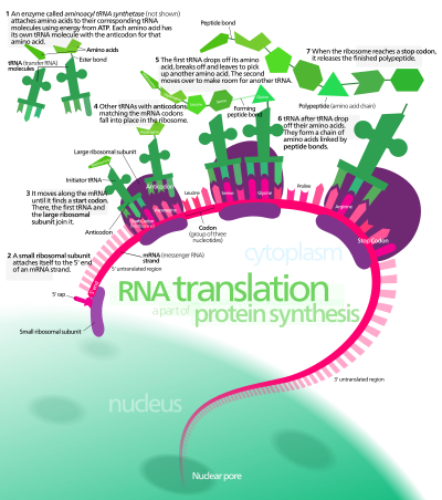
In protein synthesis, a succession of tRNA molecules charged with appropriate amino acids are brought together with an mRNA molecule and matched up by base-pairing through the anti-codons of the tRNA with successive codons of the mRNA. The amino acids are then linked together to extend the growing protein chain, and the tRNAs, no longer carrying amino acids, are released. This whole complex of processes is carried out by the ribosome, formed of two main chains of RNA, called ribosomal RNA (rRNA), and more than 50 different proteins. The ribosome latches onto the end of an mRNA molecule and moves along it, capturing loaded tRNA molecules and joining together their amino acids to form a new protein chain.[3]
Protein biosynthesis, although very similar, is different for prokaryotes and eukaryotes.
TRANSCRIPTION
In transcription an mRNA chain is generated, with one strand of the DNA double helix in the genome as a template. This strand is called the template strand. Transcription can be divided into 3 stages: initiation, elongation, and termination, each regulated by a large number of proteins such as transcription factors and coactivators that ensure that the correct gene is transcribed.
Transcription occurs in the cell nucleus, where the DNA is held. The DNA structure of the cell is made up of two helixes made up of sugar and phosphate held together by hydrogen bonds between the bases of opposite strands. The sugar and the phosphate in each strand are joined together by stronger phosphodiester covalent bonds. The DNA is "unzipped" (disruption of hydrogen bonds between different single strands) by the enzyme helicase, leaving the single nucleotide chain open to be copied. RNA polymerase reads the DNA strand from the 3-prime (3') end to the 5-prime (5') end, while it synthesizes a single strand of messenger RNA in the 5'-to-3' direction. The general RNA structure is very similar to the DNA structure, but in RNA the nucleotide uracil takes the place that thymine occupies in DNA. The single strand of mRNA leaves the nucleus through nuclear pores, and migrates into the cytoplasm.
The first product of transcription differs in prokaryotic cells from that of eukaryotic cells, as in prokaryotic cells the product is mRNA, which needs no post-transcriptional modification, whereas, in eukaryotic cells, the first product is called primary transcript, that needs post-transcriptional modification (capping with 7-methyl-guanosine, tailing with a poly A tail) to give hnRNA (heterogeneous nuclear RNA). hnRNA then undergoes splicing of introns (noncoding parts of the gene) via spliceosomes to produce the final mRNA.
TRANSLATION 

The synthesis of proteins from RNA is known as translation. In eukaryotes, translation occurs in the cytoplasm, where the ribosomes are located. Ribosomes are made of a small and large subunit that surround the mRNA. In translation, messenger RNA (mRNA) is decoded to produce a specific polypeptide according to the rules specified by the trinucleotide genetic code. This uses an mRNA sequence as a template to guide the synthesis of a chain of amino acids that form a protein. Translation proceeds in four phases: activation, initiation, elongation, and termination (all describing the growth of the amino acid chain, or polypeptide that is the product of translation).
In activation, the correct amino acid (AA) is joined to the correct transfer RNA (tRNA). While this is not, in the technical sense, a step in translation, it is required for translation to proceed. The AA is joined by its carboxyl group to the 3' OH of the tRNA by an ester bond. When the tRNA has an amino acid linked to it, it is termed "charged". Initiation involves the small subunit of the ribosome binding to 5' end of mRNA with the help of initiation factors (IF), other proteins that assist the process. Elongation occurs when the next aminoacyl-tRNA (charged tRNA) in line binds to the ribosome along with GTP and an elongation factor. Termination of the polypeptide happens when the A site of the ribosome faces a stop codon (UAA, UAG, or UGA). When this happens, no tRNA can recognize it, but releasing factor can recognize nonsense codons and causes the release of the polypeptide chain. The capacity of disabling or inhibiting translation in protein biosynthesis is used by some antibiotics such as anisomycincycloheximidechloramphenicoltetracyclinestreptomycinerythromycinpuromycin
etc.


Thursday, March 2, 2017

Success GED DNA Structure and Replication


DNA Structure and Replication: Crash Course Biology #10

In molecular biologyDNA replication is the biological process of producing two identical replicas of DNA from one original DNA molecule. This process occurs in all living organisms and is the basis for biological inheritance. DNA is made up of a double helix of two complementary strands. During replication, these strands are separated. Each strand of the original DNA molecule then serves as a template for the production of its counterpart, a process referred to as semiconservative replication. Cellular proofreading and error-checking mechanisms ensure near perfect fidelity for DNA replication.[1][2]
In a cell, DNA replication begins at specific locations, or origins of replication, in the genome.[3] Unwinding of DNA at the origin and synthesis of new strands results in replication forks growing bi-directionally from the origin. A number of proteins are associated with the replication fork to help in the initiation and continuation of DNA synthesis. Most prominently, DNA polymerase synthesizes the new strands by adding nucleotides that complement each (template) strand. DNA replication occurs during the S-stage of interphase.
DNA replication can also be performed in vitro (artificially, outside a cell). DNA polymerases isolated from cells and artificial DNA primers can be used to initiate DNA synthesis at known sequences in a template DNA molecule. The polymerase chain reaction (PCR), a common laboratory technique, cyclically applies such artificial synthesis to amplify a specific target DNA fragment from a pool of DNA.
By I, Madprime, CC BY-SA 3.0, https://commons.wikimedia.org/w/index.php?curid=2497221

DNA usually exists as a double-stranded structure, with both strands coiled together to form the characteristic double-helix. Each single strand of DNA is a chain of four types of nucleotides. Nucleotides in DNA contain a deoxyribose sugar, a phosphate, and a nucleobase. The four types of nucleotide correspond to the four nucleobases adeninecytosineguanine, and thymine, commonly abbreviated as A,C, G and T. Adenine and guanine are purine bases, while cytosine and thymine are pyrimidines. These nucleotides form phosphodiester bonds, creating the phosphate-deoxyribose backbone of the DNA double helix with the nuclei bases pointing inward (i.e., toward the opposing strand). Nucleotides (bases) are matched between strands through hydrogen bonds to form base pairs. Adenine pairs with thymine (two hydrogen bonds), and guanine pairs with cytosine (stronger: three hydrogen bonds).
DNA strands have a directionality, and the different ends of a single strand are called the "3' (three-prime) end" and the "5' (five-prime) end". By convention, if the base sequence of a single strand of DNA is given, the left end of the sequence is the 5' end, while the right end of the sequence is the 3' end. The strands of the double helix are anti-parallel with one being 5' to 3', and the opposite strand 3' to 5'. These terms refer to the carbon atom in deoxyribose to which the next phosphate in the chain attaches. Directionality has consequences in DNA synthesis, because DNA polymerase can synthesize DNA in only one direction by adding nucleotides to the 3' end of a DNA strand.
The pairing of complementary bases in DNA (through hydrogen bonding) means that the information contained within each strand is redundant. Phosphodiester (intra-strand) bonds are stronger than hydrogen (inter-strand) bonds. This allows the strands to be separated from one another. The nucleotides on a single strand can therefore be used to reconstruct nucleotides on a newly synthesized partner strand.[4]

Monday, February 27, 2017

GED Heredity and Genetics


Heredity: Crash Course Biology #9

Genetics is the study of genes—what they are, what they do, and how they work. Genes inside the nucleus of a cell are strung together in such a way that the sequence carries information: that information determines how living organisms inherit various features (phenotypic traits). For example, offspring produced by sexual reproduction usually look similar to each of their parents because they have inherited some of each of their parents' genes. Genetics identifies which features are inherited, and explains how these features pass from generation to generation. In addition to inheritance, genetics studies how genes are turned on and off to control what substances are made in a cell—gene expression; and how a cell divides—mitosis or meiosis.
Some phenotypic traits can be seen, such as eye color while others can only be detected, such as blood type or intelligence. Traits determined by genes can be modified by the animal's surroundings (environment): for example, the general design of a tiger's stripes is inherited, but the specific stripe pattern is determined by the tiger's surroundings. Another example is a person's height: it is determined by both genetics and nutrition.
Chromosomes are tiny packages which contain one DNA molecule and its associated proteins. Humans have 46 chromosomes (23 pairs). This number varies between species—for example, many primates have 24 pairs. Meiosis creates special cells, sperm in males and eggs in females, which only have 23 chromosomes. These two cells merge into one during the fertilization stage of sexual reproduction, creating a zygote. In a zygote, a nucleic acid double helix divides, with each single helix occupying one of the daughter cells, resulting in half the normal number of genes. By the time the zygote divides again, genetic recombination has created a new embryo with 23 pairs of chromosomes, half from each parent. Mating and resultant mate choice result in sexual selection. In normal cell division (mitosis) is possible when the double helix separates, and a complement of each separated half is made, resulting in two identical double helices in one cell, with each occupying one of the two new daughter cells created when the cell divides.
Chromosomes all contain DNA made up of four nucleotides, abbreviated C (cytosine), G (guanine), A (adenine), or T (thymine), which line up in a particular sequence and make a long string. There are two strings of nucleotides coiled around one another in each chromosome: a double helix. C on one string is always opposite from G on the other string; A is always opposite T. There are about 3.2 billion nucleotide pairs on all the human chromosomes: this is the human genome. The order of the nucleotides carries genetic information, whose rules are defined by the genetic code, similar to how the order of letters on a page of text carries information. Three nucleotides in a row—a triplet—carry one unit of information: a codon.
The genetic code not only controls inheritance: it also controls gene expression, which occurs when a portion of the double helix is uncoiled, exposing a series of the nucleotides, which are within the interior of the DNA. This series of exposed triplets (codons) carries the information to allow machinery in the cell to "read" the codons on the exposed DNA, which results in the making of RNA molecules. RNA in turn makes either amino acids or microRNA, which are responsible for all of the structure and function of a living organism; i.e. they determine all the features of the cell and thus the entire individual. Closing the uncoiled segment turns off the gene.
Heritability means the information in a given gene is not always exactly the same in every individual in that species, so the same gene in different individuals does not give exactly the same instructions. Each unique form of a single gene is called an allele; different forms are collectively called polymorphisms. As an example, one allele for the gene for hair color and skin cell pigmentation could instruct the body to produce black pigment, producing black hair and pigmented skin; while a different allele of the same gene in a different individual could give garbled instructions that would result in a failure to produce any pigment, giving white hair and no pigmented skin: albinismMutations are random changes in genes creating new alleles, which in turn produce new traits, which could help, harm, or have no new effect on the individual's likelihood of survival; thus, mutations are the basis for evolution.


Thursday, January 26, 2017

GED Science Biology Photosynthesis

PHOTOSYNTHESIS


Photosynthesis is a process used by plants and other organisms to convert light energy into chemical energy that can later be released to fuel the organisms' activities (energy transformation). This chemical energy is stored in carbohydrate molecules, such as sugars, which are synthesized from carbon dioxide and water – hence the name photosynthesis, from the Greek φῶςphōs, "light", and σύνθεσιςsynthesis, "putting together".[1][2][3] In most cases, oxygen is also released as a waste product. Most plants, most algae, and cyanobacteria perform photosynthesis; such organisms are called photoautotrophs. Photosynthesis is largely responsible for producing and maintaining the oxygen content of the Earth's atmosphere, and supplies all of the organic compounds and most of the energy necessary for life on Earth.[4]
Although photosynthesis is performed differently by different species, the process always begins when energy from light is absorbed by proteins called reaction centres that contain green chlorophyll pigments. In plants, these proteins are held inside organelles called chloroplasts, which are most abundant in leaf cells, while in bacteria they are embedded in the plasma membrane. In these light-dependent reactions, some energy is used to strip electrons from suitable substances, such as water, producing oxygen gas. The hydrogen freed by the splitting of water is used in the creation of two further compounds that act as an immediate energy storage means: reduced nicotinamide adenine dinucleotide phosphate (NADPH) and adenosine triphosphate (ATP), the "energy currency" of cells.
In plants, algae and cyanobacteria, long-term energy storage in the form of sugars is produced by a subsequent sequence of light-independent reactions called the Calvin cycle; some bacteria use different mechanisms, such as the reverse Krebs cycle, to achieve the same end. In the Calvin cycle, atmospheric carbon dioxide is incorporated into already existing organic carbon compounds, such as ribulose bisphosphate (RuBP).[5] Using the ATP and NADPH produced by the light-dependent reactions, the resulting compounds are then reduced and removed to form further carbohydrates, such as glucose.
The first photosynthetic organisms probably evolved early in the evolutionary history of life and most likely used reducing agents such as hydrogen or hydrogen sulfide, rather than water, as sources of electrons.[6] Cyanobacteria appeared later; the excess oxygen they produced contributed directly to the oxygenation of the Earth,[7] which rendered the evolution of complex life possible. Today, the average rate of energy capture by photosynthesis globally is approximately 130 terawatts,[8][9][10] which is about three times the current power consumption of human civilization.[11] Photosynthetic organisms also convert around 100–115 thousand million metric tonnes of carbon into biomass per year.[12][13]


Wednesday, January 18, 2017

GED ATP & Respiration Lesson

ATP and RESPIRATION


Cellular respiration is a set of metabolic reactions and processes that take place in the cells of organisms to convert biochemical energy from nutrients into adenosine triphosphate (ATP), and then release waste products.[1] The reactions involved in respiration are catabolic reactions, which break large molecules into smaller ones, releasing energy in the process, as weak so-called "high-energy" bonds are replaced by stronger bonds in the products. Respiration is one of the key ways a cell releases chemical energy to fuel cellular activity. Cellular respiration is considered an exothermic redox reaction which releases heat. The overall reaction occurs in a series of biochemical steps, most of which are redox reactions themselves. Although technically, cellular respiration is a combustion reaction, it clearly does not resemble one when it occurs in a living cell because of the slow release of energy from the series of reactions.
Nutrients that are commonly used by animal and plant cells in respiration include sugaramino acids and fatty acids, and the most common oxidizing agent (electron acceptor) is molecular oxygen (O2). The chemical energy stored in ATP (its third phosphate group is weakly bonded to the rest of the molecule and is cheaply broken allowing stronger bonds to form, thereby transferring energy for use by the cell) can then be used to drive processes requiring energy, including biosynthesislocomotion or transportation of molecules across cell membranes.


By Narayanese, WikiUserPedia, YassineMrabet, TotoBaggins - http://biocyc.org/META/NEW-IMAGE?type=PATHWAY&object=TCA. Image adapted from :Image:Citric acid cycle noi.svg|(uploaded to Commons by wadester16), CC BY-SA 3.0, https://commons.wikimedia.org/w/index.php?curid=6217701


Tuesday, January 17, 2017

GED Science PLANT CELL Lesson

PLANT CELLS 


Plant cells are eukaryotic cells that differ in several key aspects from the cells of other eukaryotic organisms. Their distinctive features include: